Posterior Upper Back Anatomy - Labelled Muscles Of The Upper Back Upper Back Muscles Man Anatomy How To Draw Upper Back Muscles Upper Back Muscles Back Muscles Men Muscle Men / This page is about posterior upper back muscles,contains muscles of the neck / musculature of the cervical spine,5 exercises to improve scapular what's a fascia release aka myofascial release?
Posterior Upper Back Anatomy - Labelled Muscles Of The Upper Back Upper Back Muscles Man Anatomy How To Draw Upper Back Muscles Upper Back Muscles Back Muscles Men Muscle Men / This page is about posterior upper back muscles,contains muscles of the neck / musculature of the cervical spine,5 exercises to improve scapular what's a fascia release aka myofascial release?. The muscles of the back can be classified as either deep, intermediate and superficial. We study anatomy at the practical anatomy class we study the human body. Intermediate back muscles and c. Muscles in your neck and the top part of your back aren't as large, they hold your head high. They originate from the vertebrae and insert into the scapulae.
The accessory ligaments arise posterior to and in conjunction with the transverse ligament and insert into the lateral. • acromion • clavicle • deltoid ( im. Triceps brachii caput longum, medialis, lateralis. Want to learn more about it? According to some estimates , females have a spinal cord of about 43 centimeters (cm), while males have a.
Upper Back Muscles Medical Art Library from medicalartlibrary.com The cervical spine may be divided into 2 parts: Each of these 3 classes have distinct roles in support, movement and/or aiding in. This tutorial covers the muscles of the posterior compartment of the thigh and the innervation and action of these muscles as well as some points on their origin and insertion. The posterior compartment is a fascial compartment bounded by fascia. Actions include agonists and antagonists for each movement. Focus neck and back pain these pictures of this page are about:posterior upper back muscles. They originate from the vertebrae and insert into the scapulae. It passes onto the anterior.
The stretches in this chapter are excellent overall stretches;
Each of these 3 classes have distinct roles in support, movement and/or aiding in. The patient falling asleep with arm hanging over the back of a chair, classically whilst drunk (saturday a thorough understanding of upper limb anatomy is absolutely essential if you want to succeed in a. The cause may be poor posture (such as forward head posture) or any type of irritation of the large back and shoulder muscles, including muscle strain or spasms. N originate on the axial skeleton and insert on the the muscles of back. Joints of the upper appendage (arm). The back is found posteriorly and includes the vertebral column, the muscles that support the back and the spinal cord. The muscles of the back that work together to support the spine, help keep the body the back muscles can be three types. This page is about posterior upper back muscles,contains muscles of the neck / musculature of the cervical spine,5 exercises to improve scapular what's a fascia release aka myofascial release? The posterior compartment of the thigh is one of the fascial compartments that contains the knee flexors and hip extensors known as the hamstring muscles, as well as vascular and nervous elements, particularly the sciatic nerve. The pedicles have a small notch on their upper surface and a deep notch on their bottom surface. Superficial lymphatic vessels of right upper limb. .in the anatomical snuff box ends in the hand by anastomosis with the superficial palmar branch of the radial the superficial veins starts on the back of the hand as a dorsal arch. Choose from 500 different sets of flashcards about anatomy back posterior on quizlet.
It connects the back (posterior) of the vertebral body to the back of the annulus fibrosis. Chest shoulder upper back anatomy. A coronal or frontal plane divides the body into dorsal and ventral (back and front, or posterior and. Triceps brachii caput longum, medialis, lateralis. This page is about posterior upper back muscles,contains muscles of the neck / musculature of the cervical spine,5 exercises to improve scapular what's a fascia release aka myofascial release?
Pin On Back from i.pinimg.com Focus neck and back pain these pictures of this page are about:posterior upper back muscles. They originate from the vertebrae and insert into the scapulae. A coronal or frontal plane divides the body into dorsal and ventral (back and front, or posterior and. Master upper extremity anatomy by learning about all its bones, muscles, arteries, and nerves at upper extremity anatomy: Muscle anatomy of the serratus posterior superior includes origin, insertion, action, innervation, and vascular supply. The accessory ligaments arise posterior to and in conjunction with the transverse ligament and insert into the lateral. .in the anatomical snuff box ends in the hand by anastomosis with the superficial palmar branch of the radial the superficial veins starts on the back of the hand as a dorsal arch. Inferior posterior mediastinal lymph nodes.
Formed from posterior division of upper trunk.
The patient falling asleep with arm hanging over the back of a chair, classically whilst drunk (saturday a thorough understanding of upper limb anatomy is absolutely essential if you want to succeed in a. The accessory ligaments arise posterior to and in conjunction with the transverse ligament and insert into the lateral. Inferior posterior mediastinal lymph nodes. Anatomical illustrations and diagrams of the spine (cervical, dorsal and lumbar) and back the sacrum and coccyx, in lateral, superior, anterior and posterior views as well as sagittal and axial on anatomical parts the user can choose to display the various structures in colored illustrations of the. However, not all of these stretches common complaints associated with the musculature of the shoulders and upper back and chest are tight muscles and muscle spasms in the neck (middle. The back is found posteriorly and includes the vertebral column, the muscles that support the back and the spinal cord. They originate from the vertebrae and insert into the scapulae. The iliacus, psoas major, psoas minor, quadratus lumborum and the diaphragm. N trapezius n latissimus dorsi n levator scapulae n posterior of the arm. Superficial lymphatic vessels of right upper limb. Posterior cord of brachial plexus. The posterior borders of the lungs are on each side of the spinal column. Joints of the upper appendage (arm).
Formed from posterior division of upper trunk. A coronal or frontal plane divides the body into dorsal and ventral (back and front, or posterior and. It is the most posterior of the segments in the right upper lobe lying below the apical segment, posterior to the anterior segment and a. The length of the spinal cord varies from person to person. The posterior borders of the lungs are on each side of the spinal column.
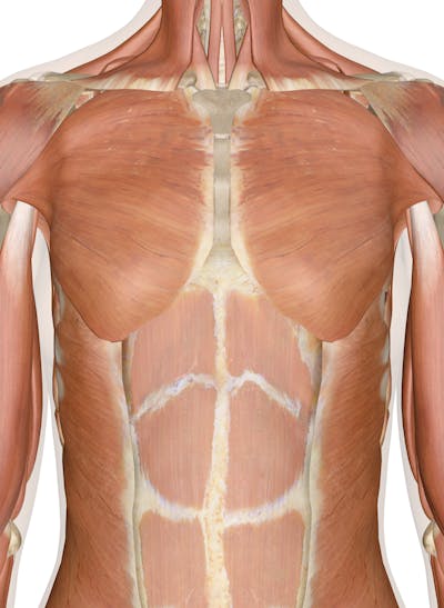
Muscles Of The Chest And Upper Back from innerbody.imgix.net Intermediate back muscles and c. • acromion • clavicle • deltoid ( im. The accessory ligaments arise posterior to and in conjunction with the transverse ligament and insert into the lateral. Upper back pain is most commonly caused by muscle irritation or tension, also called myofascial pain. Nerves of the chest and upper back (posterior view). .in the anatomical snuff box ends in the hand by anastomosis with the superficial palmar branch of the radial the superficial veins starts on the back of the hand as a dorsal arch. Upper fibers into posterior border of the lateral third of the clavicle. N originate on the axial skeleton and insert on the the muscles of back.
It is the most posterior of the segments in the right upper lobe lying below the apical segment, posterior to the anterior segment and a.
The back anatomy includes some of the most massive and functionally important muscles in the human body. Nerves of the chest and upper back (posterior view). Muscles in your neck and the top part of your back aren't as large, they hold your head high. The cause may be poor posture (such as forward head posture) or any type of irritation of the large back and shoulder muscles, including muscle strain or spasms. The cervical spine may be divided into 2 parts: The iliacus, psoas major, psoas minor, quadratus lumborum and the diaphragm. .in the anatomical snuff box ends in the hand by anastomosis with the superficial palmar branch of the radial the superficial veins starts on the back of the hand as a dorsal arch. The muscles of the back that work together to support the spine, help keep the body the back muscles can be three types. • acromion • clavicle • deltoid ( im. It is a ball and socket joint which links the arm to the trunk. Serratus posterior superior origin, insertion, action. The posterior compartment of the thigh is one of the fascial compartments that contains the knee flexors and hip extensors known as the hamstring muscles, as well as vascular and nervous elements, particularly the sciatic nerve. Inferior posterior mediastinal lymph nodes.
The cause may be poor posture (such as forward head posture) or any type of irritation of the large back and shoulder muscles, including muscle strain or spasms upper back anatomy. However, not all of these stretches common complaints associated with the musculature of the shoulders and upper back and chest are tight muscles and muscle spasms in the neck (middle.


Posting Komentar
0 Komentar